Șabloane populare
fundal sonor liber
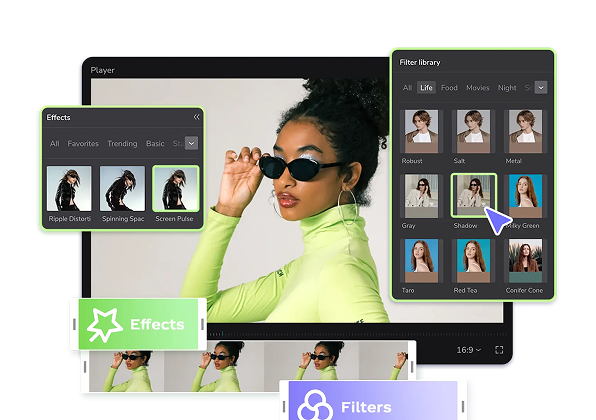
Add new video
00:03
104.2k
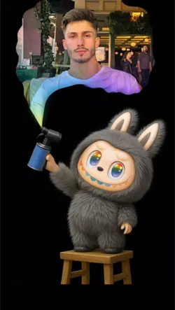
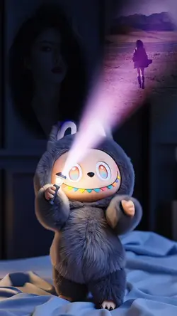
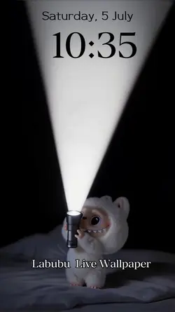
Labubu wallpaper
00:22
1.3k
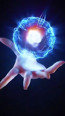
Hand power overlay
00:16
769
free shadow edit

00:10
7.8k
Bald filter

00:11
67.7k
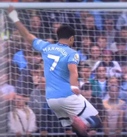
Free lamine edit
muzică de fundal hotel mp3 descărcare
fundal muzical pentru ceremonie de absolvire
muzică de fundal corporativă descărcare gratuită
descărcare muzică de fundal fericită
00:17
4.8k
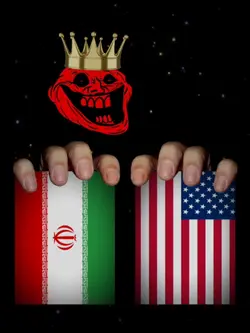
USA Iran Vs Vietnam

00:12
2.2k
Aura kid on boat

00:16
8.0k
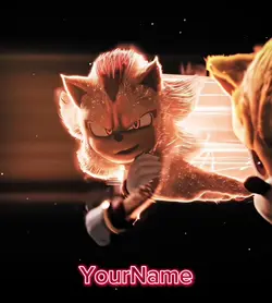
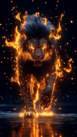
Lightning Lion

00:14
5.6k
Leao Free edit 🇵🇹

00:15
18.4k
Fire red blue intro

00:16
567
LION AI

00:09
259
Oliver!!
00:18
52.6k
Majestic ahh edit 😭
00:10
1.3k
lion fire ai
00:09
70.9k
Labubu

00:15
2.2k
Tsunami meme

00:13
165.8k
Lamine Yamal edit
00:06
11.2k
Labubu Wallpaper